Optical methods:
- fluorescence micropscopy
- bright field microscopy and similar
- imaging ellipsometry
- diffusion measurement by "fluorescence recovery after photobleaching" (FRAP)
- ns-pulsed laser setup
- and others ...
Physical or force sensitive methods:
- several different Langmuir filmbalance setups
- interfacial stress rheometer (ISR)
- surface potential measurement setup
- differential scanning calorimetry (DSC)
- surface acoustic wave (SAW) microfluidics
- and others ...
Reciprocal space methods:
- neutron reflectivity at ILL, Grenoble, France
- grazing-incidence x-ray diffraction (GID) at ESRF, Grenoble, France
- grazing-incidence x-ray out-of-plane scattering (GIXOS) at ESRF
- x-ray reflectivity at ESRF
- small- and wide-angle x-ray scattering (SAXS/WAXS) at HASYLAB/DESY, Hamburg
- and others ...
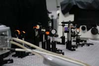
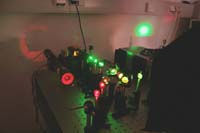
Some selected setups:
Single-dye Tracing TIRF microscope
 |  | 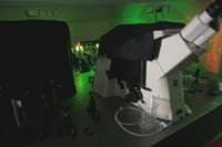 |
| To investigate diffusion processes in membranes at the smallest langth scales, we have constructed a single-dye two-color total-internal-reflection (TIRF) fluorescence microscope. Single-dye tracing (SDT) is at the limit of the technologically possible and single-molecule experiments reveal many effects hidden in classical ensemble experiments.We are especially interested in protein diffusion in cell adhesion regions, membrane junctions and similar physical conditions. | ||
Imaging Ellipsometer
 | ||
| Ellipsometry is a widely used and powerful surface sensitive method which can precisely detect thin soft-matter films of nanometer thickness. We use this method for characterization of polymer films and similar surface features. Imaging ellipsometry adds the capability to resolve lateral structures with µm resolution and nm thickness. | ||
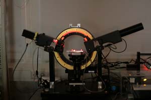
Surface Acoustic Wave (SAW) Microfluidics Workstation
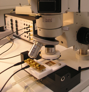 | ||
| In cooperation with Augsburg University and Advalytix, we are developing a new kind of open microfluidics system to manipulate cells in an open channel, driven by surface acoustic waves (SAW). Functionalized supported membranes in the channel can provide cell adhesion motifs and allow the investigation of cell binding under lateral stress. | ||